How Do MRI Scanners Improve Diagnostic Accuracy?

Time:2026-10-10 Author:Aria
0%

How MRI scanners improve diagnostic accuracy is a question of image quality, clinical judgment, and disciplined workflow. Unlike X-ray and CT, MRI does not use ionizing radiation. It uses magnetic fields and radiofrequency signals to separate soft tissues with remarkable detail. This contrast can reveal small lesions, torn ligaments, inflamed nerves, and early changes that may remain unclear on other scans.

The evidence is encouraging. The American College of Radiology Appropriateness Criteria identifies MRI as usually appropriate for many neurological, musculoskeletal, and oncological indications. However, “usually appropriate” does not mean universally superior. The right test depends on symptoms, timing, patient history, and the clinical question. OECD Health Statistics 2023 also shows major differences in MRI availability between countries, reminding us that access influences diagnostic pathways. More scanners do not automatically produce better diagnoses.

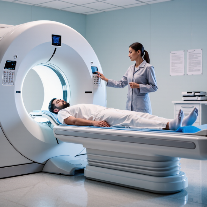
Accuracy improves when technologists select the correct sequences, adjust coils carefully, and reduce motion. A patient holding still inside the scanner can make the difference between a sharp meniscus tear and an ambiguous shadow. Radiologists then compare images with prior examinations, laboratory findings, and physical signs. The ACR Manual on MR Safety emphasizes controlled procedures, screening, and trained personnel, because safe imaging supports reliable imaging. Contrast agents may add useful information, but they require careful risk assessment, especially for patients with kidney disease.

There is room for caution. MRI can produce incidental findings, false positives, or technically limited studies. Artificial intelligence may assist interpretation, but it cannot replace accountable clinical review. Trustworthy diagnosis comes from combining validated technology, experienced professionals, and honest communication about uncertainty.
How Do MRI Scanners Improve Diagnostic Accuracy?

MRI Principles That Support Detailed Diagnostic Imaging

MRI scanners improve diagnostic accuracy by turning proton behavior into measurable signals. Most clinical images rely on hydrogen in water and fat. A strong magnetic field aligns these protons. Radiofrequency energy briefly disturbs that order. As protons recover, they release signals. The scanner records timing and strength. These differences create anatomical contrast. Small structures become easier to separate.

Timing controls what clinicians see. T1-weighted sequences often show anatomy and fat clearly. T2-weighted sequences highlight fluid, edema, and inflammation. Diffusion imaging tracks restricted water movement, supporting assessment of acute tissue injury. Gradient fields encode location, forming slices and three-dimensional views. Thin sections can reveal subtle lesions. Multiple planes add context. Yet every sequence has trade-offs. Higher detail may require longer acquisition times.

In practice, technologists reduce motion, select suitable coils, and check image quality before interpretation. Contrast agents can clarify vascularity or tissue boundaries when clinically justified. They are not always necessary. A bright signal is not a diagnosis. Artifacts, metal, breathing, and patient movement can imitate disease. Radiologists compare findings with symptoms, prior studies, and laboratory information. No scan is perfect. Even advanced imaging can miss small or unusual abnormalities. Careful protocol selection and transparent reporting make MRI evidence more dependable, though judgment remains essential.

How MRI Captures Differences Between Body Tissues

How Do MRI Scanners Improve Diagnostic Accuracy?

MRI improves diagnosis by separating tissues through their different magnetic behavior. Hydrogen atoms in water and fat respond differently inside the scanner’s magnetic field. Their signals create contrasts called T1 and T2 weighting. Muscle may appear dark, while fluid can look bright. Small changes become visible. A radiologist can then compare a lesion with nearby tissue, rather than judging shape alone.

Diffusion-weighted imaging adds another layer. It measures how freely water moves between cells. Tightly packed tissue can restrict movement and appear brighter on certain images. In the 2018 PRECISION trial, published in the New England Journal of Medicine, an MRI-based pathway detected clinically significant prostate cancer in 38% of men, compared with 26% after standard biopsy. The result supports MRI’s value, but it does not make every scan definitive.

Contrast agents can highlight blood flow and abnormal vessel patterns. However, inflammation may resemble a tumor. Motion can blur a liver edge or hide a small brain lesion. Interpretation also depends on protocol quality and reader experience. The European Association of Urology’s 2024 guideline still stresses structured reporting and clinical correlation. That matters in practice. A technically excellent image can still mislead when symptoms, laboratory results, and prior scans are ignored. MRI is powerful, but not infallible.

How Do MRI Scanners Improve Diagnostic Accuracy?

MRI distinguishes body tissues by measuring their T1 and T2 relaxation behavior. The representative values below show why tissues produce different signal intensities on different MRI sequences.

Approximate T1 and T2 relaxation times at 1.5 Tesla; values vary with field strength, sequence settings, temperature, and tissue condition.

The Role of Image Contrast in Detecting Abnormalities

MRI scanners improve diagnostic accuracy by separating tissues through image contrast. T1-weighted images make fat appear bright, while T2-weighted images highlight fluid. Diffusion-weighted imaging shows how freely water moves through tissue. Restricted movement can suggest dense cellular areas, including some tumors. Small details matter. A dark rim, uneven enhancement, or blurred boundary may change the radiologist’s interpretation.

Professional evidence supports this approach. The 2024 European Association of Urology Guidelines report pooled MRI sensitivity of about 91% for clinically significant prostate cancer, but specificity near 37%. This gap matters. Strong contrast can reveal suspicious tissue, yet it may also produce false alarms. In breast imaging, European Society of Breast Imaging recommendations cite MRI sensitivity generally above 90% for invasive cancer detection. However, background enhancement and motion can reduce practical reliability.

Contrast agents may further expose blood flow and tissue permeability. Their value depends on timing, sequence quality, and patient movement. A bright spot is not automatically dangerous. Nor is a subtle lesion harmless. Radiologists often compare multiple sequences, prior examinations, and clinical history before assigning meaning. The 2023 American College of Radiology practice guidance emphasizes standardized acquisition and interpretation, but real-world scans remain imperfect. Poor positioning, metal artifacts, or rushed protocols can weaken contrast. Better images help, but disciplined reading still decides accuracy.

Advanced MRI Techniques for More Precise Diagnosis

How Do MRI Scanners Improve Diagnostic Accuracy?

Advanced MRI techniques help clinicians examine tissue structure, blood flow, and cellular behavior with greater detail. Conventional images show anatomy clearly, but newer sequences can reveal changes before major structural damage appears. Diffusion-weighted imaging tracks water movement within tissue. Restricted movement may suggest a stroke, infection, or certain tumors. It is useful. However, it cannot provide a diagnosis alone.

Perfusion MRI measures how blood reaches different regions. Abnormal blood flow may help radiologists assess tumor activity or brain injury. Functional MRI maps changes linked to brain activity during specific tasks. This can support surgical planning near language or movement areas. Magnetic resonance spectroscopy adds chemical information, including patterns involving selected metabolites. Three-dimensional isotropic imaging also creates thin, adjustable views of complex anatomy, such as small joints or blood vessels.

Accuracy still depends on careful technique and expert interpretation. Patient movement can blur images, especially during longer examinations. Metal implants, prior surgery, and kidney conditions may also affect the imaging plan. Radiologists compare advanced findings with symptoms, examinations, and earlier scans. No sequence is flawless. A bright signal can reflect several causes, not one disease. This is where clinical judgment matters. Teams should explain uncertainty instead of overstating what an image proves. Further research may improve these tools, but responsible diagnosis still requires context, verification, and human oversight.

Clinical Factors That Influence MRI Diagnostic Accuracy

How Do MRI Scanners Improve Diagnostic Accuracy?

Clinical factors strongly influence MRI diagnostic accuracy, often as much as scanner performance. The clinical question should guide the examination protocol. A knee injury needs different sequences from suspected stroke or abdominal disease. Clear referral information helps radiologists focus on relevant anatomy and possible findings.

Patient movement remains a common source of blurred images. Pain, anxiety, confusion, and breathing difficulties can reduce image quality. Careful positioning, comfort measures, and simple instructions often make a measurable difference. Metal implants, previous surgery, and unusual anatomy also require thoughtful planning. Contrast agents may improve visibility when clinically appropriate, but kidney function, allergies, pregnancy, and current medications must be reviewed. Perfect images are uncommon. Interpretation still depends on professional judgment.

Tips: Share the exact symptoms, their duration, and previous imaging. Follow preparation instructions about eating, drinking, or medication. Stay still and report discomfort early. Ask whether motion affected the scan. Radiologists should compare earlier studies and discuss uncertain findings with the referring clinician. That conversation can prevent a technically clear image from being clinically misunderstood. Human error remains possible, especially when symptoms and images do not match. Reassessment is sometimes necessary.

FAQS

How does MRI image contrast help detect abnormalities?

Different sequences make tissues appear with different brightness levels. Fat may look bright, while fluid often stands out. Small changes become easier to compare.

What can diffusion-weighted imaging reveal?

It shows how freely water moves through tissue. Restricted movement may suggest dense cells, infection, stroke, or certain tumors. It is not a diagnosis alone.

Does a bright MRI spot always indicate disease?

No. A bright area can reflect fluid, inflammation, blood flow, or technical effects. Context matters. Bright does not mean dangerous.

Why can MRI findings produce false alarms?

High sensitivity can reveal suspicious areas that are not harmful. Background enhancement, motion, and uneven tissue can confuse interpretation. The image is not perfect.

How do contrast agents improve MRI assessment?

They can show blood flow and tissue permeability more clearly. Their usefulness depends on timing, image quality, and patient movement. Timing matters.

Which problems can reduce MRI accuracy?

Movement may blur edges and hide small details. Metal artifacts can distort nearby anatomy. Poor positioning or rushed scanning can weaken contrast.

How are advanced MRI techniques used in clinical decisions?

Perfusion imaging evaluates blood supply. Functional imaging maps activity during tasks. Spectroscopy adds chemical information. These findings support decisions, not replace judgment.

Why do radiologists compare several MRI sequences?

One sequence may show fluid, while another highlights cellular behavior or blood flow. Comparing them reduces overreliance on one signal. One image is insufficient.

What information should be combined with MRI results?

Radiologists consider symptoms, physical findings, earlier scans, and treatment history. A subtle lesion may need follow-up. Uncertainty should be stated clearly.

Can MRI provide certainty by itself?

Usually, no. Even detailed images may have overlapping appearances. Human interpretation remains important. More detail helps, but it cannot remove every doubt.

Conclusion

How MRI scanners improve diagnostic accuracy is rooted in their ability to create highly detailed images of internal body structures without using ionizing radiation. MRI principles rely on magnetic fields and radiofrequency signals to detect differences in tissue properties, allowing clinicians to distinguish muscles, organs, blood vessels, fluids, and other anatomical features. Carefully controlled image contrast makes subtle abnormalities, such as inflammation, tissue damage, or unusual growths, easier to identify and evaluate.

Advanced MRI techniques can further improve diagnostic precision by providing functional information, examining blood flow, or producing images from multiple angles and sequences. However, accurate interpretation also depends on clinical factors, including the patient’s symptoms, movement during scanning, medical history, and the quality of the imaging protocol. When these elements are combined with expert analysis, MRI can support earlier detection, clearer characterization of disease, and more informed decisions about patient care.

Aria

Aria

Aria is a dedicated marketing professional with a deep passion for innovative strategies and a keen understanding of our company's product offerings. With a wealth of experience in the industry, Aria excels at crafting engaging content that highlights the unique features and benefits of our......