An abnormal cervical screening result can bring a rush of questions. What happens next? A colposcope helps a trained clinician examine the cervix more closely. It is a magnifying instrument with a light, positioned outside the body. During the examination, the clinician may apply a mild solution to make unusual areas easier to see. If a spot needs further assessment, a small tissue sample may be taken for laboratory testing.
Why colposcopes are important for early detection comes down to seeing changes that routine screening can flag but cannot fully assess. Colposcopy can help clinicians identify areas that may need monitoring or biopsy, including changes that could develop into cervical cancer if left untreated. It does not diagnose cancer by itself, and it cannot prevent every case. Results depend on careful examination, appropriate follow-up, and clear communication between patient and clinician.
The procedure is often brief, but it can feel uncomfortable or awkward. A speculum, bright light, and unfamiliar surroundings are real details, not minor concerns. Patients should be able to ask what each step involves and discuss pain or anxiety beforehand. Follow-up matters. A normal-looking examination may not end the need for care, while an abnormal result does not automatically mean cancer. Understanding what colposcopy can—and cannot—show makes it easier to approach the next step with realistic expectations.
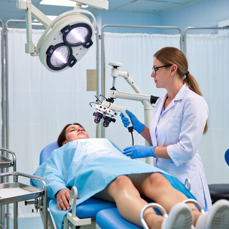
A colposcope is a lighted instrument that magnifies the cervix during a focused examination. It helps a trained clinician inspect areas that may be difficult to see with the naked eye. The patient usually lies on an examination table while a speculum gently separates the vaginal walls. The colposcope remains outside the body. No radiation is used.
During the examination, the clinician may apply diluted acetic acid or an iodine solution. These liquids can make abnormal surface changes appear white or show different staining patterns. The clinician studies the transformation zone, where many cervical abnormalities begin. If a suspicious area appears, a small biopsy may be collected for laboratory analysis. It is usually brief. A colposcope does not diagnose cancer by itself. Tissue interpretation still requires a qualified pathology professional. Inflammation, infection, or healing tissue can sometimes look concerning, so visual judgment is not perfectly objective.
The International Agency for Research on Cancer reported about 604,000 new cervical cancer cases and 342,000 deaths worldwide in 2020. These figures show why careful follow-up after an abnormal screening result matters. Colposcopy can guide the clinician toward the most relevant area for sampling. It cannot replace screening, vaccination, or reliable follow-up systems. Access also remains uneven, especially where trained providers and pathology services are limited. That limitation deserves attention. Good examination technique, clear documentation, and patient communication are essential for dependable care.
| Data Dimension | Evidence-Based Information | Why It Matters for Early Detection |
|---|---|---|
| What a colposcope is | A colposcope is a lighted binocular viewing instrument used to examine the cervix, vagina, and sometimes the vulva at enlarged magnification. | It allows a clinician to identify areas that may not be visible during a routine pelvic examination. |
| Typical magnification | Most colposcopes provide adjustable magnification, commonly ranging from approximately 3× to 15×, depending on the instrument and examination step. | Magnification helps reveal subtle changes in cervical surface patterns, blood vessels, and tissue borders. |
| Main area examined | The examination focuses on the cervix, especially the transformation zone where the outer and inner cervical tissues meet. | Most cervical precancerous changes develop in or near this biologically active area. |
| Common reason for referral | Common indications include a positive high-risk HPV test, abnormal cervical cytology, or a visible cervical abnormality. | Colposcopy provides a closer assessment after a screening result indicates an increased likelihood of cervical disease. |
| Preparation of the cervix | The clinician usually applies dilute acetic acid and may apply an iodine-based solution to highlight differences in tissue appearance. | These solutions can make suspicious areas more distinct from normal-appearing tissue. |
| Visual findings assessed | The clinician evaluates acetowhitening, iodine staining, vascular patterns, surface texture, lesion margins, and the visibility of the transformation zone. | Combining several visual signs improves the clinical assessment and helps guide targeted sampling. |
| Biopsy capability | If an abnormal area is identified, a small tissue sample may be collected for laboratory examination. An endocervical sample may also be considered when clinically appropriate. | Histopathology confirms whether abnormal cells are present and determines their severity. |
| What colposcopy can detect | It can help identify suspected cervical precancer, invasive cancer, genital warts, inflammation, and other visible tissue abnormalities. | Detection before invasive cancer develops creates an opportunity for timely monitoring or treatment. |
| Typical examination setting | Colposcopy is commonly performed in an outpatient clinical setting, with the patient positioned similarly to a routine pelvic examination. | An outpatient procedure can provide diagnostic evaluation without requiring hospital admission in most cases. |
| Time required | The visual examination often takes about 10 to 20 minutes, although the total visit may be longer if biopsies, documentation, or counseling are required. | The relatively short procedure supports efficient follow-up after an abnormal screening result. |
| Patient experience | The speculum examination may cause pressure or discomfort. A biopsy can cause brief pinching or cramping, and light spotting may occur afterward. | Clear preparation and communication can reduce anxiety and support completion of recommended follow-up. |
| Diagnostic limitation | Colposcopy is a visual assessment and cannot alone provide a definitive diagnosis in every case. Biopsy results and clinical history may be necessary. | Recognizing its limitations helps prevent both overdiagnosis and missed disease. |
| Follow-up after examination | Follow-up depends on the examination findings, biopsy results, HPV status, age, pregnancy status, and applicable clinical guidelines. | Risk-based follow-up ensures that confirmed lesions receive appropriate surveillance or treatment while lower-risk findings are not overtreated. |
Note: Colposcopy is a diagnostic examination and should be interpreted by a qualified healthcare professional together with cervical screening results and, when indicated, tissue pathology.
Why Are Colposcopes Important for Early Detection?
When Colposcopy Is Recommended After Screening
An abnormal cervical screening result does not automatically mean cancer. It may show cell changes or detect high-risk human papillomavirus (HPV), which can raise concern when it persists. A clinician may recommend colposcopy when results suggest a higher chance of significant cell changes. Not every positive HPV test needs one. The decision depends on the specific result, age, and screening history, as well as current clinical guidance.
During colposcopy, a clinician examines the cervix through a magnifying instrument. If an area looks unusual, they may take a small tissue sample for laboratory testing. The procedure can feel uncomfortable, and that deserves plain acknowledgment. A referral can be worrying, but it is a closer look, not a cancer diagnosis. Many biopsy results show changes that need monitoring or treatment rather than cancer.
Ask what finding prompted the referral and when follow-up should happen. If the recommendation is unclear, request an explanation before the appointment. Screening records matter: previous results can change the next step. Guidance can also vary by individual circumstances. A missed appointment may delay clarification, so contact the clinic if timing or preparation is difficult.
A colposcope gives the clinician a close, well-lit view of the cervix without entering the body. During an examination, the patient lies on an exam table while a speculum gently opens the vagina. The clinician looks through the instrument from outside, checking the cervix for changes in color, surface, or blood-vessel patterns. Small details matter.
A mild solution, often dilute acetic acid, may be applied to make some abnormal areas easier to see. They can appear temporarily white against the surrounding tissue. This change helps guide the examination, but it does not prove that cancer or a precancerous condition is present. Irritation and harmless changes can look unusual too. The view is only one part of the assessment.
If a spot looks concerning, the clinician may take a small biopsy for laboratory review. Colposcopy helps target that sample, rather than relying on appearance alone. It can also help determine whether follow-up or further testing is appropriate. Still, the view is not the whole story. Lighting, anatomy, inflammation, and the examiner’s experience can affect what is visible. Some abnormal cells are subtle, and a normal-looking area does not always settle every concern. That deserves care. Results should be interpreted alongside screening history and biopsy findings, with the next steps explained clearly to the patient.
An abnormal screening result is a signal, not a diagnosis. Colposcopy lets a trained clinician inspect the cervix under magnification, apply a solution that helps highlight unusual areas, and take targeted biopsies. The view is not perfect. Inflammation, bleeding, or a partly hidden transformation zone can make assessment harder, so visual impressions alone cannot confirm precancer.
The International Agency for Research on Cancer’s Global Cancer Observatory estimated 662,301 new cervical cancer cases and 348,874 deaths worldwide in 2022. These figures show why timely follow-up matters, but not every abnormal screening result requires a biopsy. Clinicians consider screening history, age, HPV results, and colposcopic findings when deciding next steps.
ASCCP’s Colposcopy Standards recommend biopsying visible acetowhite areas when appropriate. A pathologist examines the tissue to identify and grade changes, helping clinicians distinguish benign findings from precancer or cancer.
Clear follow-up instructions matter, because a missed appointment can delay decisions.
A colposcope gives a clinician a magnified view of the cervix, often after an abnormal screening result. Bright light helps reveal areas that may look different, such as patches that change color after a solution is applied. The examination can guide a small biopsy from a specific spot. It does not, by itself, confirm cancer. A microscope examination of sampled tissue is usually needed to identify cell changes.
Colposcopy shows surface details, not the whole story. A normal-looking cervix cannot rule out every problem, and the procedure does not assess all conditions of the uterus or ovaries. Some changes are subtle, too high in the cervical canal to see clearly, or absent on the day of the exam. That limitation can be unsettling. Follow-up depends on screening results, biopsy findings, and personal medical history; a clinician can explain what each result means.
Tips: Ask what prompted the referral and whether a biopsy may be taken. Bring questions, and mention medicines or bleeding concerns beforehand. If you feel nervous, say so; the clinician can explain each step and pause when needed. Keep recommended follow-up appointments, even if the exam looks reassuring.
No. It may show cell changes or high-risk HPV, but it is not a cancer diagnosis. The next step depends on your age, results, and screening history.
Colposcopy may be recommended when results suggest a higher chance of significant cell changes. Not every positive HPV test leads to one.
A clinician examines the cervix through a magnifying instrument. A solution may help highlight unusual areas. The view can be imperfect.
If an area looks unusual, the clinician may take a small tissue sample. A pathologist checks it to identify and grade cell changes.
It can be. A biopsy may cause brief discomfort or light spotting. Ask the clinic what to expect.
Results may show benign changes, precancer, or cancer. Some mild changes are monitored; higher-grade changes often need treatment.
Previous results can affect the recommended next step. Ask what finding prompted your referral and when follow-up should happen.
No. Inflammation, bleeding, or a partly hidden area can make assessment harder. Visual impressions alone cannot confirm precancer.
Ask the clinic to explain the reason and preparation. If timing is difficult, contact them. A missed appointment can delay decisions. It is worth asking why this step fits your case.
A colposcope is a magnifying instrument that helps a clinician examine the cervix more closely, usually after an abnormal screening result or when other findings suggest that further assessment may be useful. It does not enter the body; instead, it provides a clearer view of cervical tissue so areas that look unusual can be examined and, when appropriate, sampled for testing.
Why colposcopes are important for early detection is that they can help clinicians locate and assess abnormal-looking tissue before deciding on next steps. The examination may guide whether a biopsy or follow-up is needed and help inform diagnosis and treatment planning. However, a colposcope alone cannot confirm the cause of an abnormal area or predict how it will develop. Its findings must be considered alongside screening results, biopsy results when taken, and the person’s overall clinical circumstances.

Suffolk Medical