Digital X-ray systems are reshaping routine imaging in hospitals, clinics, and mobile radiography units. In 2026, buyers are comparing detector sensitivity, workflow speed, cybersecurity, service support, and total ownership costs. The best system is not always the most expensive one. It must fit patient volume, room design, staff skills, and clinical priorities.
What are the benefits of digital X-ray systems? The answer begins with practical evidence. Images appear within seconds, allowing radiographers to check positioning before the patient leaves. Electronic storage also reduces film handling and supports faster image sharing with authorized clinicians. The International Atomic Energy Agency emphasizes optimization and dose management in digital radiography, not simply faster exposure. That distinction matters. A clear image with an unnecessarily high dose is not a success.
Industry forecasts from Grand View Research and MarketsandMarkets report continued growth in the digital X-ray market through the decade. Their analyses point to stronger demand for wireless detectors, automated workflow tools, and advanced image processing. However, market forecasts are not clinical guarantees. Vendor claims need careful testing. The U.S. Food and Drug Administration also stresses performance, safety, and appropriate use when evaluating medical imaging equipment.
There are limitations. Detectors can be damaged. Software may require training. Integration problems can delay reporting. Costs can rise when maintenance contracts are overlooked. This guide examines leading 2026 systems through an evidence-based lens, combining technical specifications, user experience, regulatory considerations, and real-world workflow value. Better decisions require more than a glossy brochure.
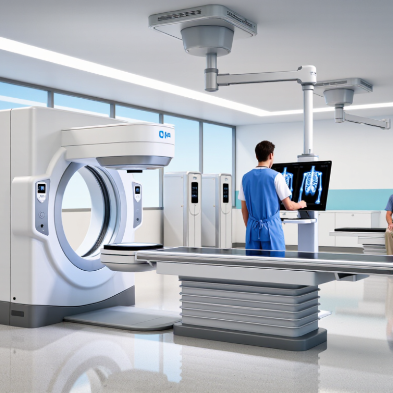
Digital X-ray uses flat-panel detectors to convert transmitted radiation into an electronic image. The image appears within seconds, rather than waiting for film processing. This matters in busy emergency rooms, where a misplaced cassette can delay clinical decisions.
The World Health Organization estimates that about 3.6 billion diagnostic X-ray examinations occur worldwide each year. That scale makes detector performance, workflow, and radiation management important. The International Atomic Energy Agency recommends optimizing exposure for each patient, not simply lowering every setting. Modern systems can support this process through exposure indicators, image review, and repeat-analysis tools.
Flat-panel detectors also improve positioning flexibility. A technologist can capture a chest image beside a bed, with fewer patient transfers. Digital images can enter a hospital archive quickly and support remote review. Yet speed is not the same as accuracy. A bright, sharp image may still hide motion, rotation, or poor patient positioning. That assumption deserves challenge.
Tips: Check detector size, pixel spacing, durability, and cleaning procedures before purchase. Ask for independent dose and repeat-rate data, not only vendor demonstrations. Train staff to interpret exposure indicators correctly. Review rejected images every month. Small workflow errors can become expensive habits.
The benefits depend on disciplined use. Equipment alone cannot guarantee safer examinations or better diagnosis. WHO data shows the scale; daily clinical practice determines the quality.
Digital radiography (DR) systems can convert X-ray energy into visible images within seconds. A detector captures the radiation after it passes through the patient’s body. Its internal sensors create electrical signals from different exposure levels. Software then processes these signals into a grayscale image.
The image appears on a workstation almost immediately. Technologists can check positioning while the patient remains nearby. This helps reduce repeated exposures caused by unclear anatomy or movement. For example, a chest image may show lung fields, ribs, and soft tissue in one screen. Doctors can adjust brightness, contrast, and magnification without developing film. That speeds clinical review and supports more informed decisions.
The process is quick, but not flawless. Poor positioning, motion, or incorrect exposure can still reduce image quality. A highly processed image may look acceptable while hiding subtle details. Experienced staff must review the raw data, patient history, and clinical request together. Routine quality checks also matter, including detector calibration and dose monitoring. In practice, the fastest image is not always the most useful one. Careful technique remains essential, even when the screen responds instantly.
2026 Best Digital X-Ray Systems: What Are the Benefits?
Patient dose optimization is central to safe digital radiography. Under IAEA safety principles, every examination should be justified and optimized. FDA requirements also support reliable equipment performance, quality control, and patient protection. The goal is not simply the lowest exposure. It is a diagnostic image with an appropriate dose.
In daily practice, technologists can reduce unnecessary exposure through accurate positioning, tight collimation, and suitable exposure settings. Automatic exposure control can help, but it cannot correct poor patient positioning. Pediatric patients need especially careful protocols. Their smaller bodies and longer expected lifetimes require thoughtful technique selection. Small details matter.
Review the exposure index after each examination. Compare repeat images, rejected studies, and local diagnostic reference levels. These records can reveal overexposure, motion errors, or inconsistent training. A clinical physicist should support periodic testing and equipment evaluation. Written procedures should reflect IAEA guidance and applicable FDA requirements.
A modern detector may improve efficiency. It does not replace professional judgment. No protocol is perfect. Patient size, anatomy, and clinical urgency can change the correct approach. I have seen dose-saving efforts fail when staff chase a number without checking image quality. Regular audits, open reporting, and practical refresher training create safer habits. The best system is therefore part of a wider safety program, not a standalone purchase.
Representative adult effective-dose estimates show that examinations vary substantially in radiation exposure. Digital radiography supports dose optimization through appropriate exposure selection, image-quality review, repeat-rate reduction, and protocol control. The IAEA and FDA emphasize justification and optimization; these values are estimates, not patient dose limits or universal diagnostic reference levels.
Source: RadiologyInfo.org, “Radiation Dose in X-Ray and CT Exams,” developed by the RSNA and ACR; safety principles: IAEA Radiation Protection of Patients and U.S. FDA medical X-ray guidance.
The 2026 best digital X-ray systems should support more than fast image capture. Their clinical value depends on reliable DICOM communication. DICOM allows images and patient details to move between compatible devices. This reduces manual entry and helps limit identification errors. Technologists can send studies directly to PACS for review, storage, and controlled sharing. That workflow matters during busy emergency shifts.
PACS gives radiologists access to current and previous examinations in one workspace. Comparing a new chest image with an older study can reveal subtle changes. It also reduces delays caused by searching through separate archives. However, integration is not automatic. Incorrect patient data, weak network performance, or poor training can still interrupt care. The system needs routine testing and clear staff procedures.
Lower repeat-image rates are another practical benefit. Better detectors can capture useful detail with fewer failed exposures. Image previews help technologists check positioning before the patient leaves. This may reduce avoidable repeats, patient discomfort, and workflow congestion. Still, no system fixes every positioning mistake. A rushed examination can produce an unusable image. Clinical teams should review repeat data regularly, identify common causes, and adjust protocols carefully. Small improvements become meaningful when measured across months.
Choosing a 2026 digital X-ray system requires more than comparing image samples.
In practical evaluations, DQE shows how efficiently a detector uses radiation to produce useful image detail. A higher DQE can support lower exposure, but only when positioning and processing are consistent. Review DQE at relevant dose levels, not just one impressive laboratory figure. Small differences may matter during repeated chest or orthopedic examinations.
Detector size affects positioning, workflow, and patient coverage.
A larger detector can reduce retakes for bedridden patients, while a smaller panel may be easier to handle. Weight matters too. Ask staff to lift and place the detector repeatedly. That simple test often reveals problems missed in brochures. MTBF provides a useful reliability signal, but it is not a promise of uninterrupted service. Request service records, warranty conditions, replacement times, and preventive maintenance requirements. I have seen purchasing teams overvalue specifications and underestimate daily handling. That mistake deserves reflection.
Tips:
Calculate total cost across five to seven years. Include detectors, software updates, calibration, training, batteries, repairs, downtime, and possible retakes. Compare installation needs and service coverage in writing. Do not treat the lowest purchase price as the lowest cost. Ask for acceptance testing and image-quality verification by qualified personnel. Keep a short trial log from real examinations; it may expose workflow friction that a sales demonstration hides.
A flat-panel detector converts transmitted radiation into an electronic image within seconds. No film processing is needed. Speed helps busy emergency rooms.
About 3.6 billion diagnostic X-ray examinations occur worldwide each year. Efficient detectors can support useful images at optimized exposure levels. More expensive does not always mean better.
Yes. A portable detector can capture a chest image beside a bed. This may help patients who cannot stand or move safely. Positioning still needs careful checking.
No. Motion, rotation, and poor positioning can remain hidden. A beautiful image may still be clinically weak. That assumption needs questioning.
Review DQE, detector size, pixel spacing, weight, durability, and cleaning procedures. Check performance at relevant dose levels. One laboratory figure is not enough.
MTBF offers a useful reliability signal, but it does not promise continuous operation. Request service records, replacement times, warranty terms, and maintenance requirements. Ask staff to handle the panel repeatedly.
Estimate costs across five to seven years. Include calibration, software updates, batteries, repairs, training, downtime, and repeat examinations. The lowest purchase price may not be the lowest total cost.
Train staff to interpret exposure indicators correctly. Review rejected images every month and record repeat patterns. Small workflow errors can become expensive habits. Equipment alone cannot fix them.
Digital X-ray systems are expected to remain a central imaging solution in 2026, using flat-panel detectors to transform X-rays into detailed digital images within seconds. With billions of examinations performed worldwide each year, this technology supports faster workflows, easier image storage, and more efficient clinical decision-making. A key question for healthcare providers is: What are the benefits of digital X-ray systems? The answer includes improved image accessibility, reduced processing time, and better support for patient-focused care.
Modern systems can help optimize radiation dose while following recognized safety principles and regulatory requirements. Their compatibility with DICOM and PACS enables seamless image transfer, review, and archiving across departments. Advanced image quality may also reduce the need for repeat examinations, improving efficiency and patient comfort. When comparing systems, facilities should evaluate detective quantum efficiency, detector dimensions, reliability measured by mean time between failures, workflow features, service support, and total cost of ownership. A balanced assessment helps organizations select a system that delivers dependable performance, long-term value, and consistent diagnostic support.

Suffolk Medical